A proton (in human tissues) produce magnetic field that transmit electromagnetic energy. The strength of the energy depends on density of the proton in the tissues and through the first and second process of scan. Each point in space that received and transmits the signal has unique frequency that can construct image possible.
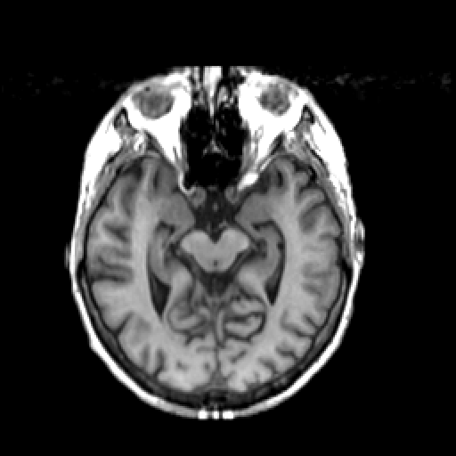
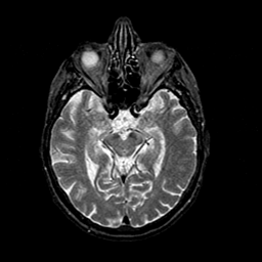
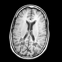
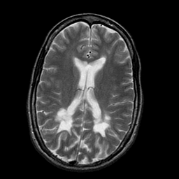
MRI (magnetic resonance imaging) When protons (here brain protons) are placed in a magnetic field, they become capable of receiving and then transmitting electromagnetic energy. The strength of the transmitted energy is proportional to the number of protons in the tissue. Signal strength is modified by properties of each proton's microenvironment, such as its mobility and the local homogeneity of the magnetic field. MR signal can be "weighted" to accentuate some properties and not others.
When an additional magnetic field is superimposed, one which is carefully varied in strength at different points in space, each point in space has a unique radio frequency at which the signal is received and transmitted. This makes constructing an image possible. It represents the spatial encoding of frequency, just like a piano.
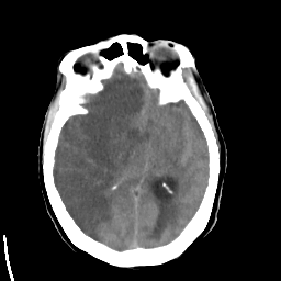
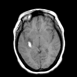
A simplified tabulation of tissue image characteristics:
Normal tissue
|
| |||
| dense bone | dark | dark | bright |
| air | dark | dark | dark |
| fat | bright | bright | dark |
| water | dark | bright | dark |
| brain | "anatomic" | interm. | interm. |
2. Bright means high density/high attenuation of x-rays, dark means low.
3. Grey matter appears gray, white matter white.
Abnormal tissue
|
| MR-T1 | MR-T2 | xray-CT | enhancement1 |
| subacute | ||||
| bleed | bright2 | no | ||
| dark3 | ||||
| dark4 | acute |
2. Unless very fresh or very old.
3. Unless calcified.
4. Often isodense.

No comments:
Post a Comment